Machine Learning for Automated Mitral Regurgitation Detection from Cardiac Imaging
Ke Xiao, Erik Learned-Miller, Evangelos Kalogerakis, James Priest & Madalina Fiterau
MICCAI 2023 Paper, MR vs non-MR video, Patch Selection video, Github repo
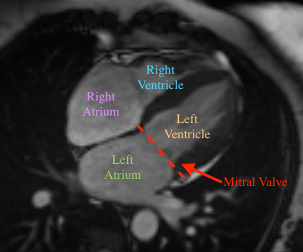
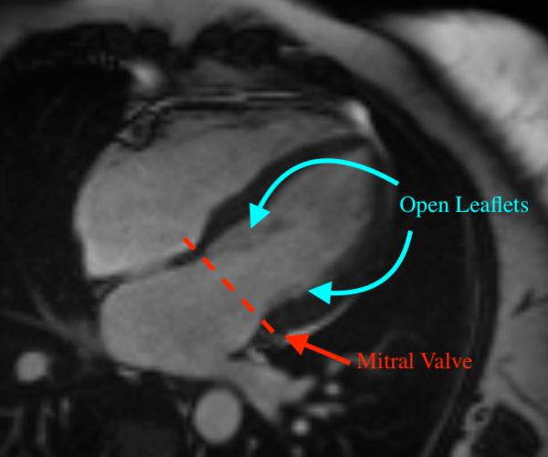
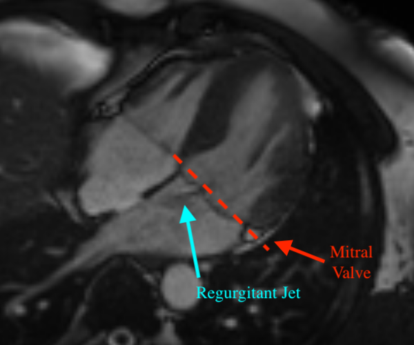
Mitral regurgitation (MR) is a heart valve disease with potentially fatal consequences that can only be forestalled through timely diagnosis and treatment. Traditional diagnosis methods are expensive, labor-intensive and require clinical expertise, posing a barrier to screening for MR. To overcome this impediment, we propose an automated five stage pipeline named Cardio-vascular magnetic resonance U-Net localized Self-Supervised Predictor (CUSSP). CUSSP operates on cardiac magnetic resonance (CMR) imaging slices of the 4-chamber view of the heart. It uses standard computer vision techniques and contrastive models to learn from large amounts of unlabeled data, in conjunction with specialized classifiers to establish the first ever automated MR classification system using CMR imaging sequences. Evaluated on a test set of 179 labeled — 154 non-MR and 25 MR — sequences, CUSSP attains an F1 score of 0.69 and a ROC-AUC score of 0.88, setting the first benchmark result for detecting MR from CMR imaging sequences.
Challenges:
- Requires multiple video frames, and tracking blood flow.
- The regurgitant jet can be faint in the video, and thus difficult to detect.
- Blood flow has high turbulence
Segmentation
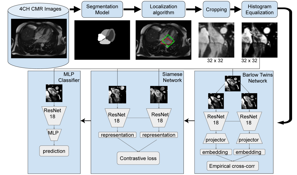
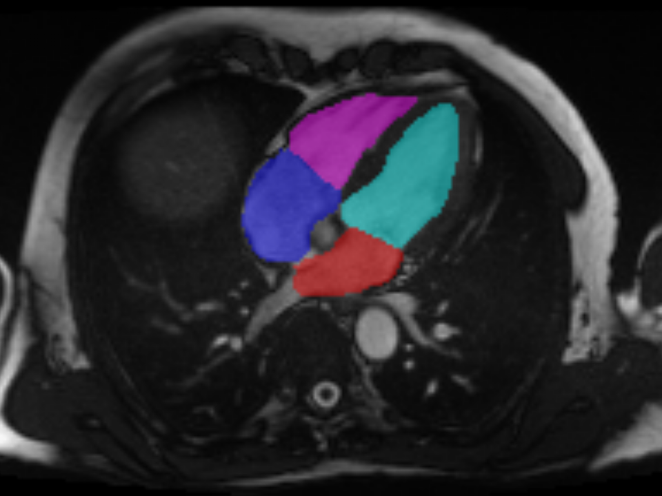
- Segmentation for CMR views including long-axis 2-chamber (LAX-2CH) view, 4-chamber (LAX-4CH) view and short-axis (SA) view.
- For patch selection:
Semantic segmentation of 4CH CMR imaging sequences. - For tabular data:
Semantic segmentation of LAX-2CH, LAX-4CH and SA CMR imaging
sequences (to estimate the cardiac structure and functions in Table 1).
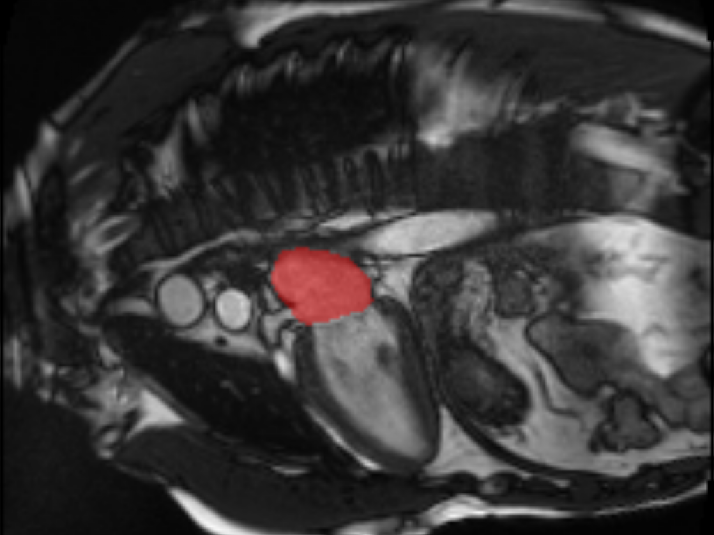
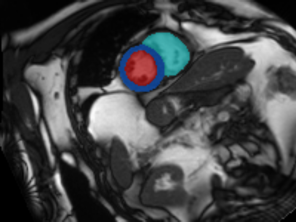
view imaging data and the short axis (right) CMR imaging data
| Left Atrium | Right Atrium | Left Ventricle | Right Ventricle |
| Vol Max (mL) Vol Min (mL) Stroke Vol (mL) Ejection Fraction (%) | Vol Max (mL) Vol Min (mL) Stroke Vol (mL) Ejection Fraction (%) | End Systolic Vol (mL) End Diastolic Vol (mL) Stroke Vol (mL) Ejection Fraction (%) Cardiac Output (L/min) Mass (g) | End Systolic Vol (mL) End Diastolic Vol (mL) Stroke Vol (mL) Ejection Fraction (%) |
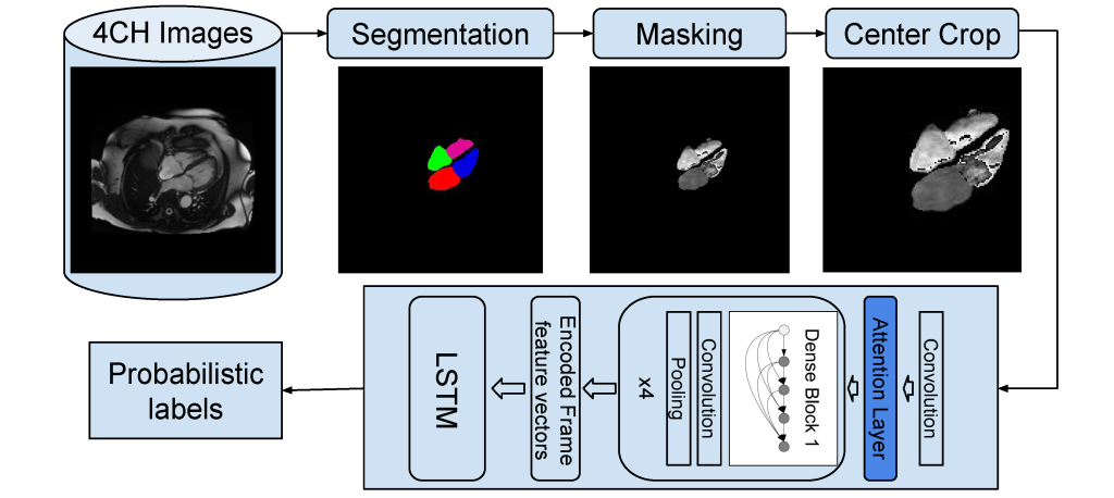
Our Prior Attempts: Random Forests and CNN-LSTM
- Random forest (RF) classifier trained on the tabular heart measurements shown in Table 1.
- A deep learning model using a CNN-LSTM pipeline, shown in Figure 3.
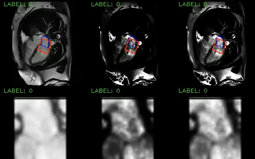
Patch Selection
- Localization: localizing the region in the heart where mitral regurgitation occurs.
- Equalization: increasing contrast for the regurgitant jet.
- Selection: cropping the patch for the ML system.
CUSSP Pipeline Stages
- CUSSP pipeline: (1) segmentation, (2) localization, (3) cropping, (4) equalization, and (5) prediction
- Prediction steps: (i) Barlow-Twins network, (ii) Siamese network, (iii) Multilayer perceptron (MLP).